
What Is Muscular Dystrophy?
Muscular dystrophy refers to a group of degenerative genetic disorders that lead to the breakdown of muscular tissue during the progression of the disease. Muscles weaken over time as tissues are replaced with fatty deposits. Eventually, an individual with muscular dystrophy may require the use of a wheelchair due to the loss of muscle mass. It can also result in the development of scoliosis, a condition in which the spine curves sideways.
Muscular Dystrophy in Children
There are many different types of muscular dystrophy. Depending on the type, onset may occur anywhere from infancy to beyond middle age. Duchenne and Becker are the two most common types of muscular dystrophies that develop in early childhood, and both typically occur in boys.
While children are usually diagnosed between the ages of 3 and 6, early detection plays an essential role in slowing the progression of muscular degeneration.
Symptoms
Signs of muscular dystrophy in children may include:
- Walking on tiptoes
- Clumsiness
- Difficulty jumping or climbing
- Weakness in the upper arms and legs
- Tripping and falling often
- Having trouble opening and closing their eyes
- Weakness of the facial muscles
Causes
Muscular dystrophy is a genetic disorder caused by abnormalities in the genes responsible for healthy tissue development. It may be passed down from one or both parents. While the occurrence of this disease in family members increases the risk of inheriting it, muscular dystrophy may also appear in individuals with no family history of the disease.
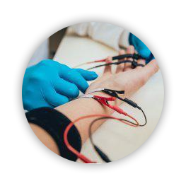
EMG Testing
One highly effective method for diagnosing muscular dystrophy is through intramuscular electromyography testing, also known as EMG. It is a test that detects muscular abnormalities by measuring the electrical impulses in skeletal muscle tissue in response to stimulation.
During the test, an instrument known as an electromyograph measures electrical impulses detected by one or more needles inserted into the patient’s muscle. These needles act as conductors or electrodes. The electromyograph converts the resulting impulses into a visual record which is then displayed in a graph-like format on the screen of an oscilloscope.
Other Common Tests
In addition to performing an EMG, other tests used to diagnose muscular dystrophy include:
- Electrocardiogram (EKG or ECG): Electrodes attached to the skin of the torso and limbs detect arrhythmias and dysrhythmias, or irregular heartbeat patterns. An EKG records the heart’s electrical impulses and can be used to identify weakness in cardiac musculature.
- Blood tests: Blood samples taken from the patient can be used to discover genetic abnormalities
- Muscle biopsy: Small portions of muscular tissue are surgically removed and inspected under a microscope.
Treatment
Unfortunately, there is no cure for muscular dystrophy. It is a life-long condition that progresses over time. However, with early detection and treatment, you can delay the progression of muscular degeneration. Treatments may include:
- Medication
- Splints and braces to support skeletomuscular structure
- Physical therapy
- Orthotic devices and mobility aids, such as walkers and crutches
- Surgery
The Arizona Institute of Neurology and Polysomnography offers cutting-edge intramuscular electromyography testing. If your child is displaying early signs of muscular dystrophy, utilizing this technology can play a critical role in early detection and getting your child the treatment they need.
